Book Your 2D Echo Test in Navi Mumbai
Get accurate heart scans from certified partner labs in Kharghar. Schedule your cardiac ultrasound appointment online with transparent pricing.
View Test PricesA Simple, 3-Step Booking Process
We connect you with trusted Navi Mumbai diagnostic centers seamlessly.
1. Find Your Test
Choose the 2D Echo test and see pricing from our partner labs in Navi Mumbai.
2. Book via WhatsApp
Click to book your preferred test. Our team will help you schedule at a convenient lab.
3. Get Your Report
Visit the lab for your test and receive your digital report, typically within 24 hours.

Understanding the 2D Echo Test
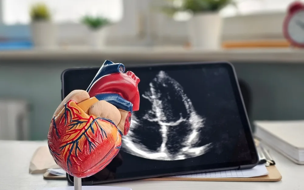
A 2D Echocardiogram is a safe, non-invasive ultrasound of the heart. It provides crucial information about your heart’s health without any radiation. A skilled technician uses a probe (transducer) with gel on your chest to capture real-time images of your heart’s muscles, chambers, and valves.
What Does a 2D Echo Report Reveal?
- Pumping Function (Ejection Fraction): Measures how effectively your heart pumps blood. A normal EF is typically 50-70%.
- Heart Size & Wall Thickness: Detects enlargement or thickening of heart walls, which can indicate conditions like hypertension or cardiomyopathy.
- Valve Function: Checks for leaky (regurgitation) or narrowed (stenosis) heart valves.
- Structural Abnormalities: Identifies congenital defects or damage from a previous heart attack.
Who Should Consider a 2D Echo Test?
Your doctor may recommend this test if you experience symptoms like chest pain, shortness of breath, palpitations, or have risk factors like high blood pressure or diabetes.
Transparent 2D Echo Test Pricing in Navi Mumbai
Affordable and clear pricing for high-quality cardiac diagnostic services.
Standard 2D Echo Test
Market Price: ₹2500
₹1899 /-
Direct Booking Discount: Save ₹601
- Complete Heart Structure & Chamber Evaluation
- Heart Valve Function Assessment
- Report by Certified Cardiologist
2D Echo + Colour Doppler
Market Price: ₹4000
₹2999 /-
Direct Booking Discount: Save ₹1001
- Includes Standard 2D Echo evaluation
- Colour Doppler Study for Blood Flow Analysis
- Detailed Ejection Fraction & Pressure Gradient Report
What Our Users Say
Hear from others who have used our platform to book their diagnostic tests.
“The booking process on Henotic Diagnostics was so simple. I found a lab near me in Kharghar and scheduled my 2D Echo in minutes. Highly efficient!”
– Sunil K.
“I appreciated the transparency. All the information about the partner lab was available upfront. A very professional and trustworthy platform.”
– Priya S.
“Great experience! The platform made it easy to find a convenient appointment time without having to make multiple phone calls. Will use it again.”
– Rohan M.
Visit Our Kharghar Center
Henotic Diagnostics
Address: Second floor, Millennium Empire, Business Park, Plot No 47, D Mart Rd, Sector 15, Kharghar, Navi Mumbai, Maharashtra 410210
Call Us: 088793 27184, 093728 53584
Website: henoticdiagnostics.com/
Our Network of Certified Partner Labs


Find a Partner Near You in Mumbai & Navi Mumbai
Search our full network of licensed labs, hospitals, and specialty clinics.
No Partner Labs Found
Please try adjusting your search terms or selecting a broader area.
Frequently Asked Questions
Is Henotic Diagnostics a lab or a clinic?
Henotic Diagnostics is neither. We are a healthcare services platform. We partner with a network of independent, certified laboratories to make it easier for you to book and manage your diagnostic appointments.
Is the 2D ECHO test painful or safe?
The test is completely safe and painless. It uses ultrasound waves (no radiation) and is even safe during pregnancy. You might feel mild pressure on your chest from the probe.
How long does the 2D Echo test take?
A standard 2D Echo test typically takes about 20 to 30 minutes to complete.
Do I need to fast before the test?
No, fasting is not required for a standard 2D Echo test. You can eat, drink, and take your regular medications unless specifically advised otherwise by your doctor.
Ready to Schedule Your Appointment?
Take the next step in managing your health. Book your 2D Echo Test in Navi Mumbai through our simple and secure platform.
Call to Book: 08879327184